This scan should be ideally performed between 12 weeks 5 days and 13 weeks 6 days of your pregnancy. Women’s Imaging conducts a detailed risk assessment for your baby in accordance with the Fetal Medicine Foundation. As well as checking that your baby is growing well and confirming your due date the main aim of the scan includes:
- To see if there are any structural abnormalities. Some can already be identified or suspected at this stage so a normal appearing scan is very reassuring for you.
- To check your uterus and ovaries to ensure they won’t be an issue during the pregnancy.
- To confirm the growth of the pregnancy and the due date.
- To check where the placenta is lying, where the umbilical cord is in relation to the placenta and if there is sufficient fluid around the baby.
Risks
Ultrasound is safe to use throughout your pregnancy.
Sometimes we need to do a vaginal scan. If you are allergic to latex prior to the vaginal scan or you don’t know then a latex-free cover will be used on the probe.
Occasionally there is some discomfort from probe pressure on a full bladder or from the vaginal probe manipulation. If this is extremely painful please let us know.
Benefits
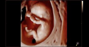
You will be able to see all of your developing baby
We are able to take some important measurements which allows us to give you an accurate risk assessment for your baby.
What is an ultrasound?
An ultrasound scan uses high-frequency sound waves to create images of the inside of your body and baby. Sound waves are used instead of radiation which makes them safe.
What do I need to do to have a risk assessment for my baby?
You will need to have a blood test done before coming to Women’s Imaging for your ultrasound scan. Your doctor will organise this for you. Ideally you should have your 1st Trimester bloods/ Maternal Serum done at 11 weeks gestation. Your doctor will organise a form for you.
We like to scan you between 12.5 and 13.6 weeks of pregnancy.
Why is a 12-14 weeks scan different at Women’s Imaging?
We offer the most sensitive and comprehensive risk assessment for your baby between 12.5-13.6 weeks which includes pre-eclampsia and fetal growth restriction. We are currently the only practice in Tasmania that does this. Traditional chromosomal abnormalities such as Down’s Syndrome continue to be assessed. Our sonographers are trained to the highest standards to perform these specialist measurements of you and your baby. They hold Certificates in Competence from the Fetal Medicine Foundation which is recognised by the Royal Australian and New Zealand College of Obstetricians and Gynaecologists.
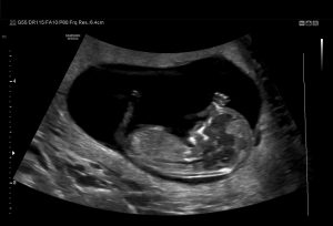
We measure:
- Your baby’s nasal bone
- The thin layer of fluid at the back of the baby’s neck (the nuchal translucency)
- Your blood pressure (we take 2 measurements from both arms)
- The blood flow between you and your baby
We also examine:
- Your blood results
- Your age
- Your weight
- Whether you have had any previous problems in pregnancies (if relevant).
When you choose to have your First Trimester scan with us, you can be confident that the risk assessment results you receive are the best available in obstetric scanning today.
Women’s Imaging provides a comforting and safe environment with the latest state of the art equipment to ensure that you have the best possible care and experience.
What happens on the day?
When you arrive for your scan you will be asked to fill out a form about your pregnancy to date.
You will then have your blood pressure taken on both arms and your height and weight will be measured if you are not sure. Sometimes your measurements are taken before you have your scan and sometimes after.
 When the sonographer takes you through to the scanning room you will be asked to lie on the table and expose your tummy. A towel will be tucked into your pants to limit spread of the gel onto your clothes.
Clear gel is applied to your tummy and the sonographer moves the probe over your tummy recording images. The clear gel is water-soluble so it won’t stain your clothes. It’s just a little sticky!
Please come with a full bladder which will make it easier to obtain images of your baby. A full bladder pushes the uterus away from your lower pelvis thereby making it easier for us to see and scan your baby.
We will take measurements of your baby and check your placenta and ovaries.
Occasionally a vaginal scan is also performed to give us a better view of your baby. You will be able to watch the entire scan on our wall-mounted plasma screen monitors!
Once the scan is completed the sonographer will leave the room. All of your measurements including your blood pressure and blood results will be reviewed by our radiologist or on-site obstetrician. Results will be sent to your referring doctor and they will give them to you at your next appointment.
You will be given your expected due date on the day. You will receive a text message within 24 hours of your appointment with a link to access your images through our online portal. Your images will be available to view within 3 days of your appointment.
How long will it take?
The 12-14 week scan takes approximately 60 minutes.
(Please allow some extra time in your schedule when you come to see us for a 12-14 week scan. Occasionally we have to recalculate your measurements and this can take a few extra minutes)
Important things to know
- This ultrasound scan is best performed between 12 weeks and 13 weeks 6 days gestation.
- The scan usually takes 45 to 60 minutes depending on the position of the baby.
- It is important that you have 1st Trimester bloods/Maternal Serum done 7 full working days before you come for your scan. Your doctor will organise this for you.