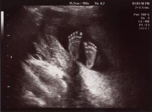
This is the main ultrasound scan that is obtained during the pregnancy.
By 20 weeks most of your baby has developed such that screening of the organs is possible to assess for abnormalities. This scan can be done anytime between 18-22 weeks of your pregnancy but ideally around the 20 week mark.
The scan can take a minimum of 45 minutes and sometimes up to an hour.
At this scan the different parts of the baby will be measured to confirm that there has been satisfactory growth in line with that is expected from the due date.
Multiple images of different organs will be taken to confirm that the development is progressing as expected.
While most of the imaging is targeted to the baby, the placenta, umbilical cord and amniotic fluid are also imaged. The blood flow to the uterus will be assessed with Doppler measurements.
Often the sex of the baby can be identified. Please let us know if you would like to know this.
Occasionally not all of the baby can be assessed at one examination. This may be because of the position of the baby. A repeat examination is sometimes required. This will be part of the original examination fee and a booking will be made for you prior to leaving the practice. Ideally we will try and complete the entire scan on the day but sometimes bub doesn’t want to show us everything! Your baby can be comfy in one spot for the entire scan!
What will happen on the day
When you arrive for your scan you will be asked to fill out a form about your pregnancy to date.
The sonographer will then take you through to the scanning room.
You will be asked to lie on the table and expose your tummy. A towel will be tucked into your pants to limit spread of the gel onto your clothes.
Clear gel is applied to your tummy and the sonographer moves the probe over your tummy recording images.
Please come with a partially filled bladder which will make it easier to obtain images of your baby.
Ultrasound at 18 weeks
We will take measurements of your baby and check your placenta and your cervix.
Occasionally a vaginal scan is also performed to give us a better view of your baby or if you have a low-lying placenta. You will be able to watch the entire scan on our wall-mounted plasma screen monitors!
In this scan, we like your baby to move around a little (not alot!) so we can scan everything we want to see.
All of our sonographers are accredited with the Fetal Medicine Foundation. Each sonographer’s scanning style is different. Some like to talk you through the scan whilst others like to stay silent as that helps them concentrate on your baby. The sonographer will explain this to you before the scan. Getting a detailed look at your baby’s anatomy is our number one priority.
Once the scan is completed, the sonographer will leave the room and the results of the scan will be reviewed by our radiologist or on-site obstetrician. If there is any cause for concern, they will come and speak to you on the day. Occasionally your baby is just too comfortable in one spot and we are unable to complete the full scan. This means that you may need to come back again for us to review the parts we haven’t seen.
You will receive a text message within 24 hours of your appointment with a link to access your images through our online portal. Your images will be available to view within 3 days of your appointment.
Risks
Ultrasound has been used for many years in pregnancy and is regarded as safe to you and your baby.
Sometimes we need to do a vaginal scan. If you are allergic to latex prior to the vaginal scan or you don’t know then a latex-free cover will be used on the probe.
Occasionally there is some discomfort from probe pressure on your bladder or from the vaginal probe manipulation. If this is extremely painful please let us know.
Unfortunately a normal ultrasound does not guarantee a normal baby. There are regrettably many problems that cannot be determined through imaging. The baby still has 20 weeks to continue to grow and develop.
Benefits
You will be able to see your developing baby
We are able to assess the heart and brain in great detail.
If there is anything that we are concerned about from the scan we will inform you on the day. This is very reassuring for you and your baby.
What is an Ultrasound scan?
An ultrasound scan uses high-frequency sound waves to create images of the inside of the body and your baby. Sound waves are used instead of radiation which makes them safe.
Why have a 20 week scan at Women’s Imaging?
Our sonographers are trained to the highest standards to perform these specialist measurements of you and your baby. They hold Certificates in Competence from the Fetal Medicine Foundation which is recognised by the Royal Australian and New Zealand College of Obstetricians and Gynaecologists. We take detailed measurements of your baby. All of our scans are monitored by our in-house radiologist or obstetrician.
How long will it take?
The 20 week scan takes approximately 45 minutes but please give yourself lots of time. Occasionally we can’t see/scan everything because of your baby’s position or the position of your placenta. Your own BMI also affects how well we can see your baby on ultrasound. Sometimes you have to rebook for another day so we can complete the scan fully.
How should I prepare for the scan?
You should arrive for your scan with a partially filled bladder. Remember to bring your referral form with you.
Important things to know
- The scan will take about 45 minutes.
- Often the sex of the baby can be identified, please let us know if you would like to know this.
- Our highly accredited sonographers work hard at checking all of your baby’s anatomy including the heart and brain. Please be aware that some sonographers like to scan for the most part in silence as it helps them concentrate on your baby. They will explain to you their scanning style at the start of the scan.