The Scan
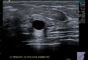
Ultrasound uses pulsed high-frequency soundwaves to produce images of the internal structure of the breast. This is in real time such that movement and blood flow can also be demonstrated. Ultrasound has no known harmful effects on humans.
Ultrasound is used for further assessment of lumps or abnormalities demonstrated on mammography. It is the first imaging method used for assessment of breast problems in patients under 35 years of age.
What happens on the day?
 The examination will take up to 30 minutes to perform. You will be asked to remove clothing from your upper body and to lie on your back, possibly slightly rotated with the assessment-side arm raised above your head. This position enables the breast to be flat and uniform thickness on the chest wall. Clear gel is applied to the breast and the sonographer moves the probe (transducer) over the breast, recording images. Once the imaging is complete the gel is removed.
Risks
There is usually no discomfort from this procedure, however some pressure or minor pain may be experienced when the transducer passes over a tender area.
During the procedure, the sonographer will leave the room to discuss the images with the radiologist. The radiologist may re-scan areas of interest. This is normal practice.
If you have any questions please ask the sonographer or radiologist.
Benefits
This scan uses no radiation.
Our on-site breast radiologist reviews the images before you leave the practice.
The radiologist will come and speak to you on the day of your scan and may rescan you also if there are any concerns.
Things you should know
- There is usually no discomfort from this study.
- The examination takes up to 30 minutes to perform.
- Our on-site Breast Radiologist will review the images before you leave the practice